We received three Tanner crab (Chionoecetes bairdi)hemolymph samples from Pam Jensen (NOAA) yesterday. From her email to Steven:
Hi Steven, I am sending: tube #1 crab 3859/3656: 300 ul blood + 1300 ul RNAlater
tube #2 crab 3665/3873: 300 ul blood + 1300 ul RNAlater tube #3 crab 3665/3873: 200 ul blood + 1400 ul RNAlater
The tubes hold max of 1600 ul. Will know on Sun or Mon if either crab is infected w Hematodinium.
Tracking info to follow. Pam
Samples were stored at 4C O/N.
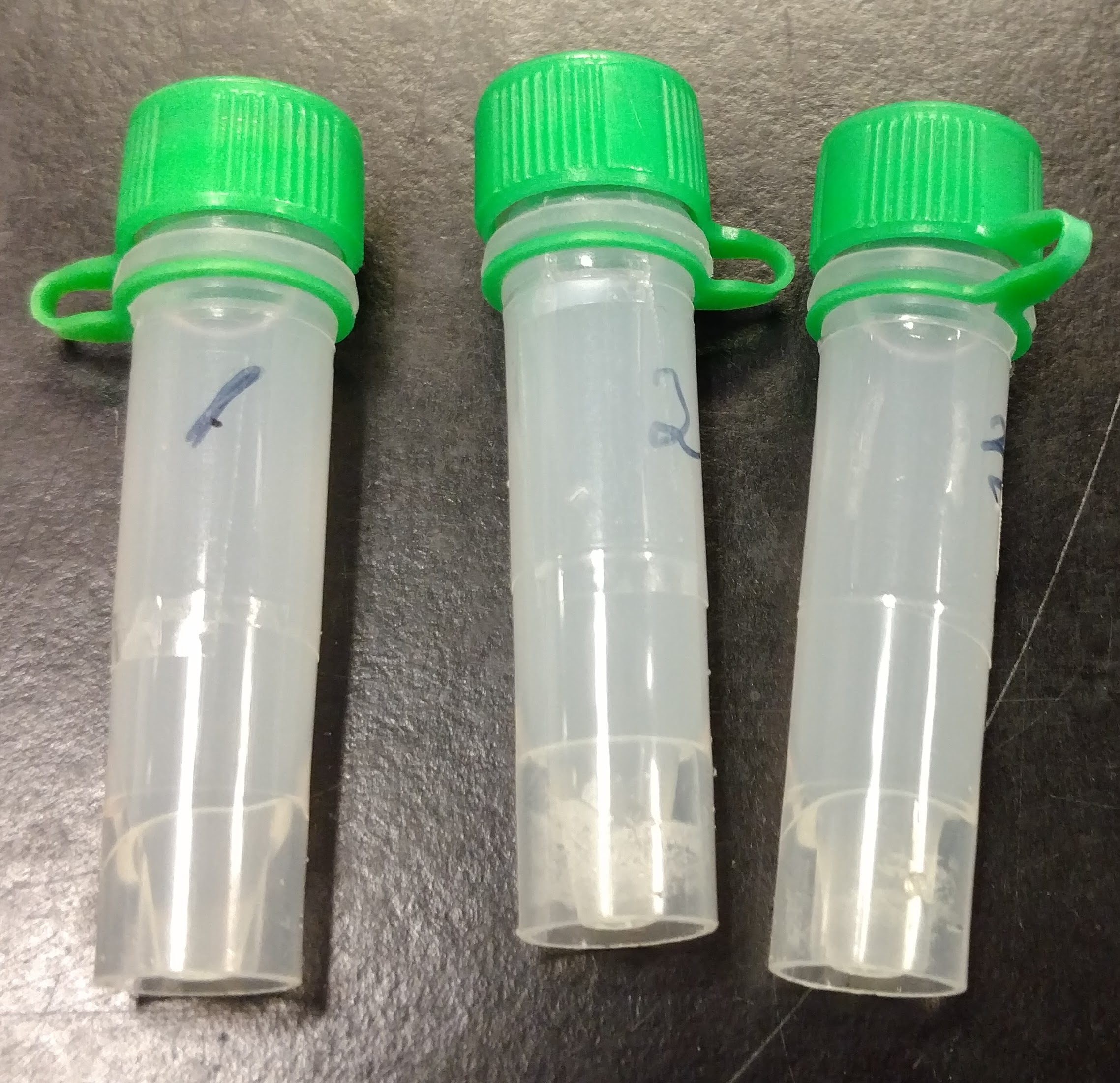
Here’s what the samples looked like before processing:
 (http://owl.fish.washington.edu/Athaliana/20171107_RNA_isoaltion_crab_01.jpg)
The samples are extremely cloudy. I’m not sure if this is expected.
Processed samples using RNAzol RT (MRC) according to the manufacturer’s protocol for Total RNA Isolation.
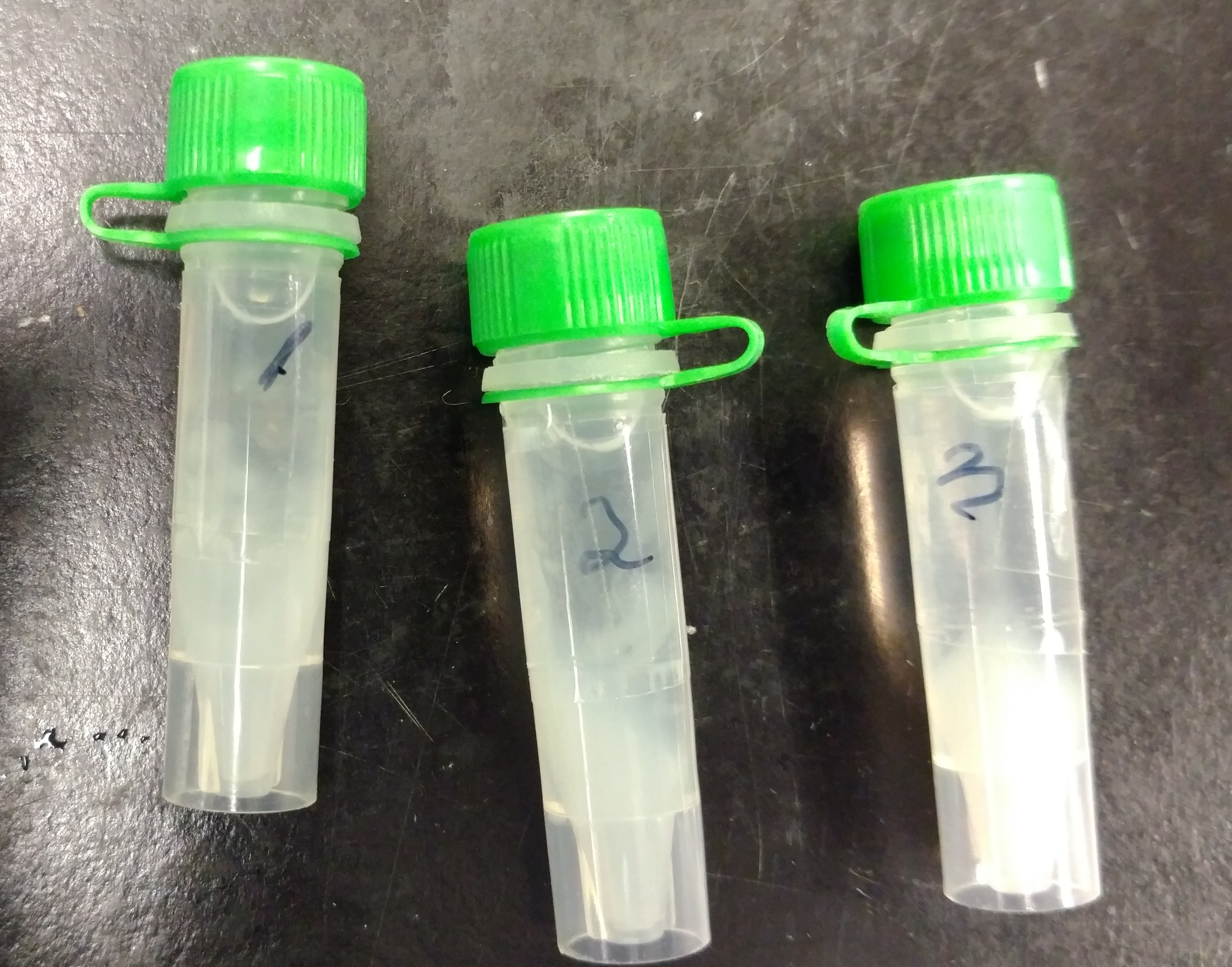
Pelleted samples at 5000g for 5 mins and the samples looked like this:
 (http://owl.fish.washington.edu/Athaliana/20171107_RNA_isoaltion_crab_02.jpg)
Decided to pellet samples for an additional 10mins. The pellet was more compact. Transferred supernatant to clean tube, since it seemed to contain “debris” (maybe cells?). Processed pellet with RNAzol RT. Brief rundown of procedure (all steps at room temp):
Transferred supe to clean tube.
Added 1mL RNAzol RT to pellet and mixed by repeated pipetting (solution was cloudy and slightly viscous).
Added 400uL of 0.1% DEPC-treated H2O and mixed vigorously by hand.
Incubated for 10mins.
Centrifuged 12,000g for 15mins.
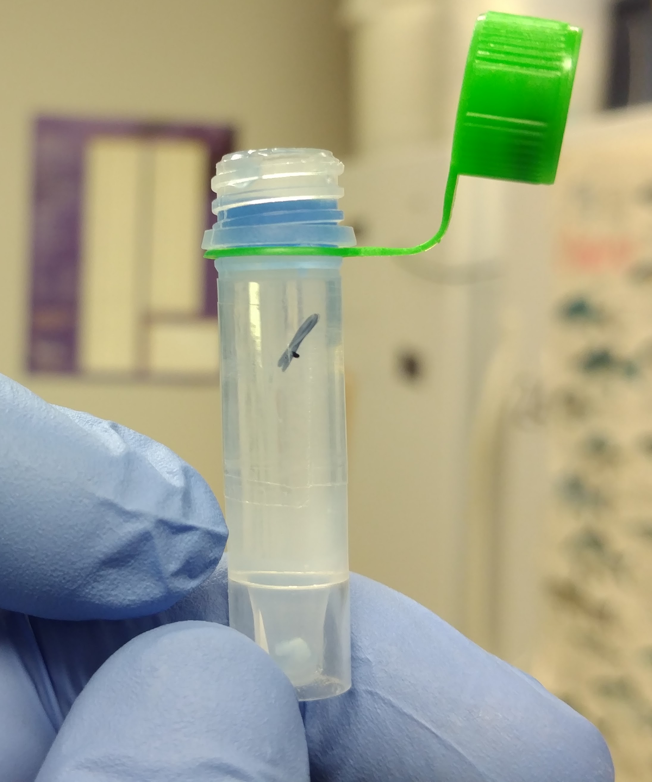
Samples looked like this:
 (http://owl.fish.washington.edu/Athaliana/20171107_RNA_isoaltion_crab_03.jpg)
This is not normal. Usually the supernatant is the clear portion, while the blue layer is below that.
Transferred 750uL of the clear portion to clean 1.7mL tube.
Added equal volume of isopropanol, mixed by inversion. Appeared to be a very high amount of genomic DNA precipitation visible in the tube.
Incubated for 10mins.
Centrifuged 12,000g, 15mins.
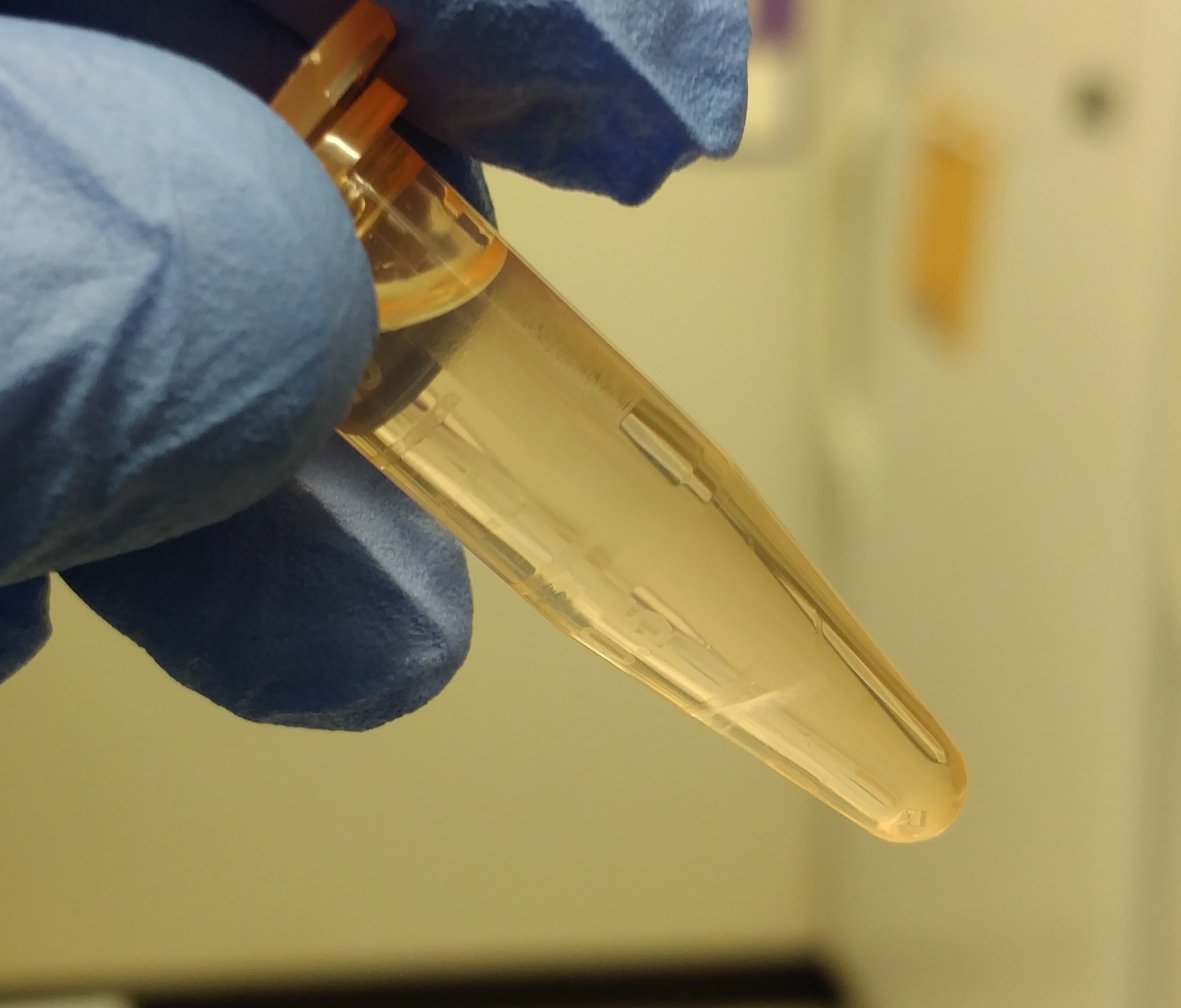
Samples looked like this:
 (http://owl.fish.washington.edu/Athaliana/20171107_RNA_isoaltion_crab_04.jpg)
It appears that the nucleotides (the white interphase) are suspended on a “cushion” of higher density solution, instead of pelleted at the bottom of the tube.
Removed/discarded higher density solution, leaving the white layer on the bottom of the tube.
Centrifuged 12,000g, 15mins.
Discarded supe.
Washed pellet with 75% ethanol.
Centrifuged 8,000g, 3mins.
Repeated Steps 12, 13, & 14, 1x.
Discarded ethanol.
Resuspended RNA in 50uL 0.1% DEPC-treated H2O. Pellets did not solubilize on their own. I dispersed the pellets by repeated pipetting (P200). Remaining insoluble material was pelleted (12,000g, 30s) and supernatant was transferred to a new 1.6mL tube.
RNA was quantified using the Qubit 3.0 and the Qubit HS RNA Assay. Used 5uL of each sample.
Results:
20171107_qubit_tanner_crab_hemo (Google Sheet)
Sample ID Conc. (ng/uL) Total Yield (ng)| 3859/3656 | 0.44 | 22 |
| 3665/3873 | 1.66 | 83 |
| 3665/3873 | 2.04 | 102 |
Interestingly, both samples from the same crab had similar/decent yields.
Samples were labeled and stored at -80C in Shellfish RNA Box #6