INTRO
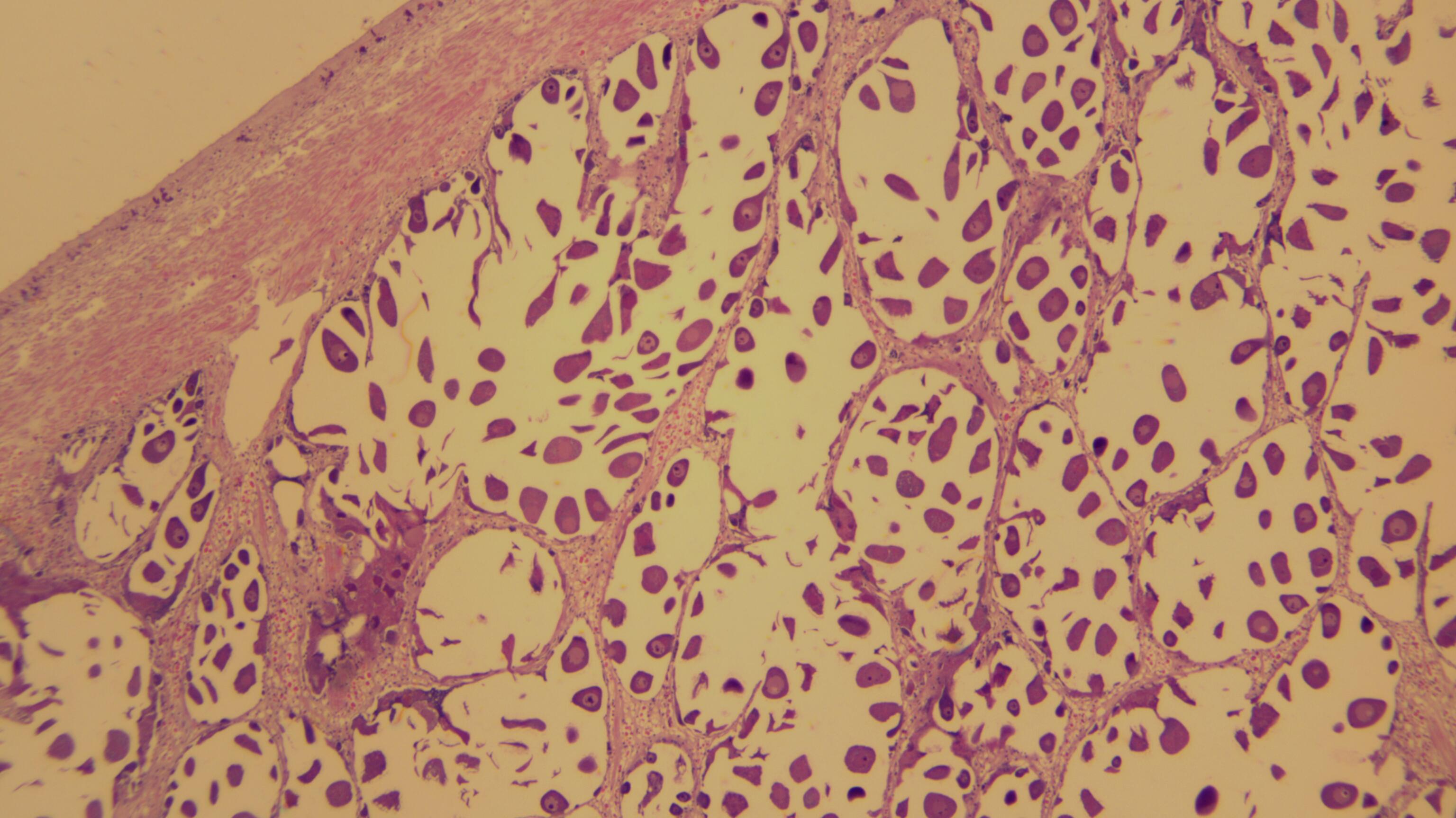
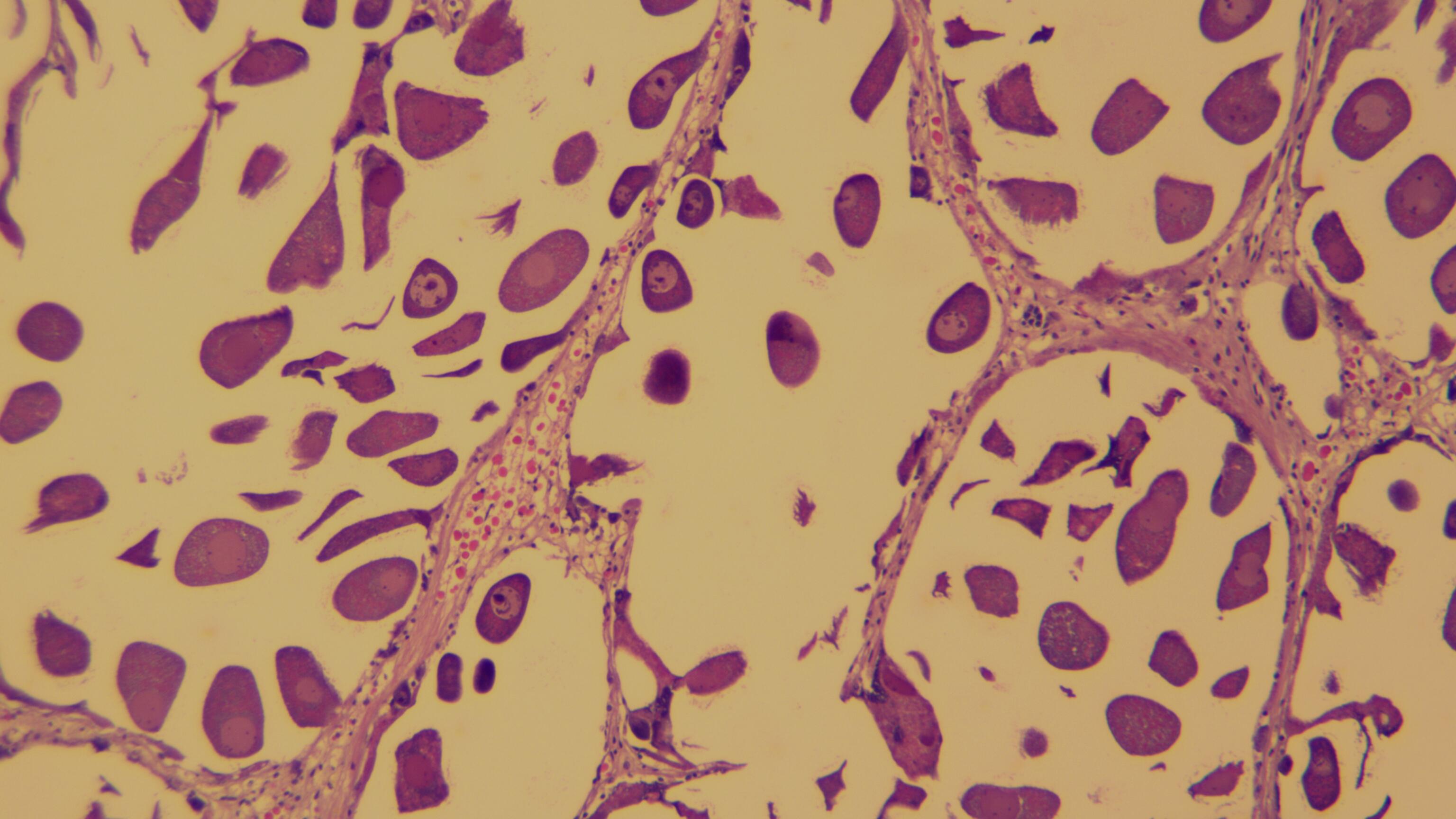
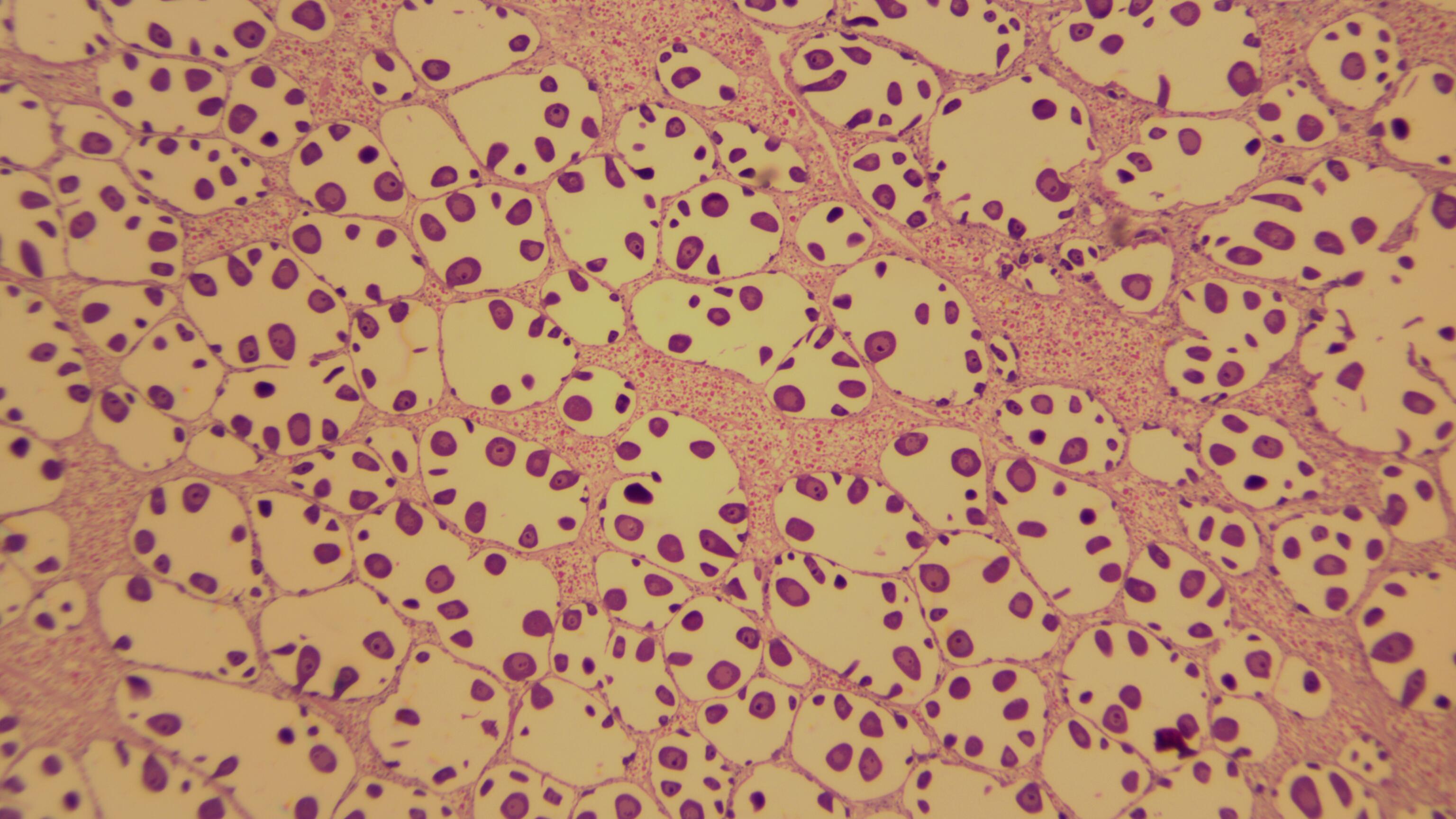
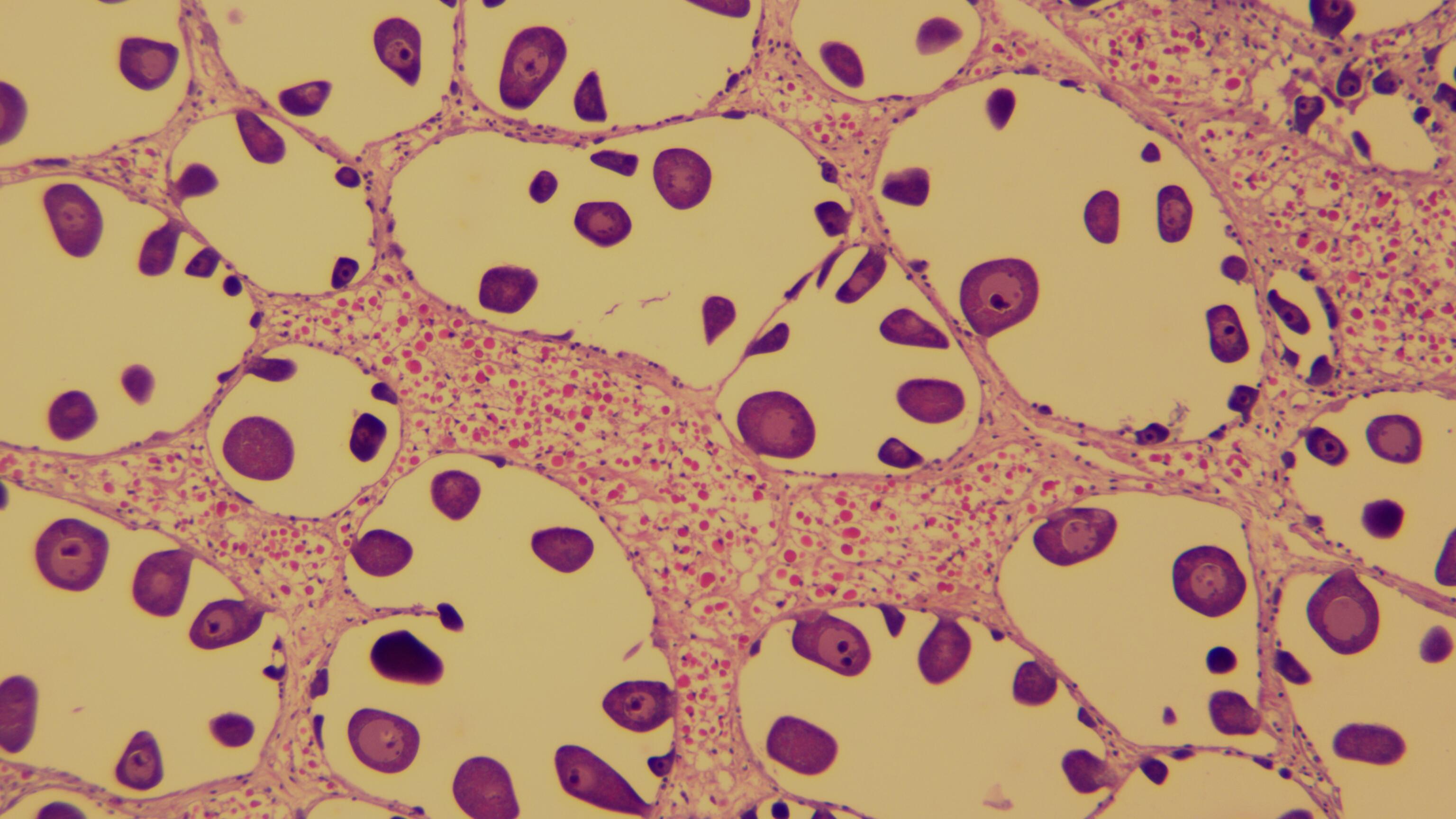
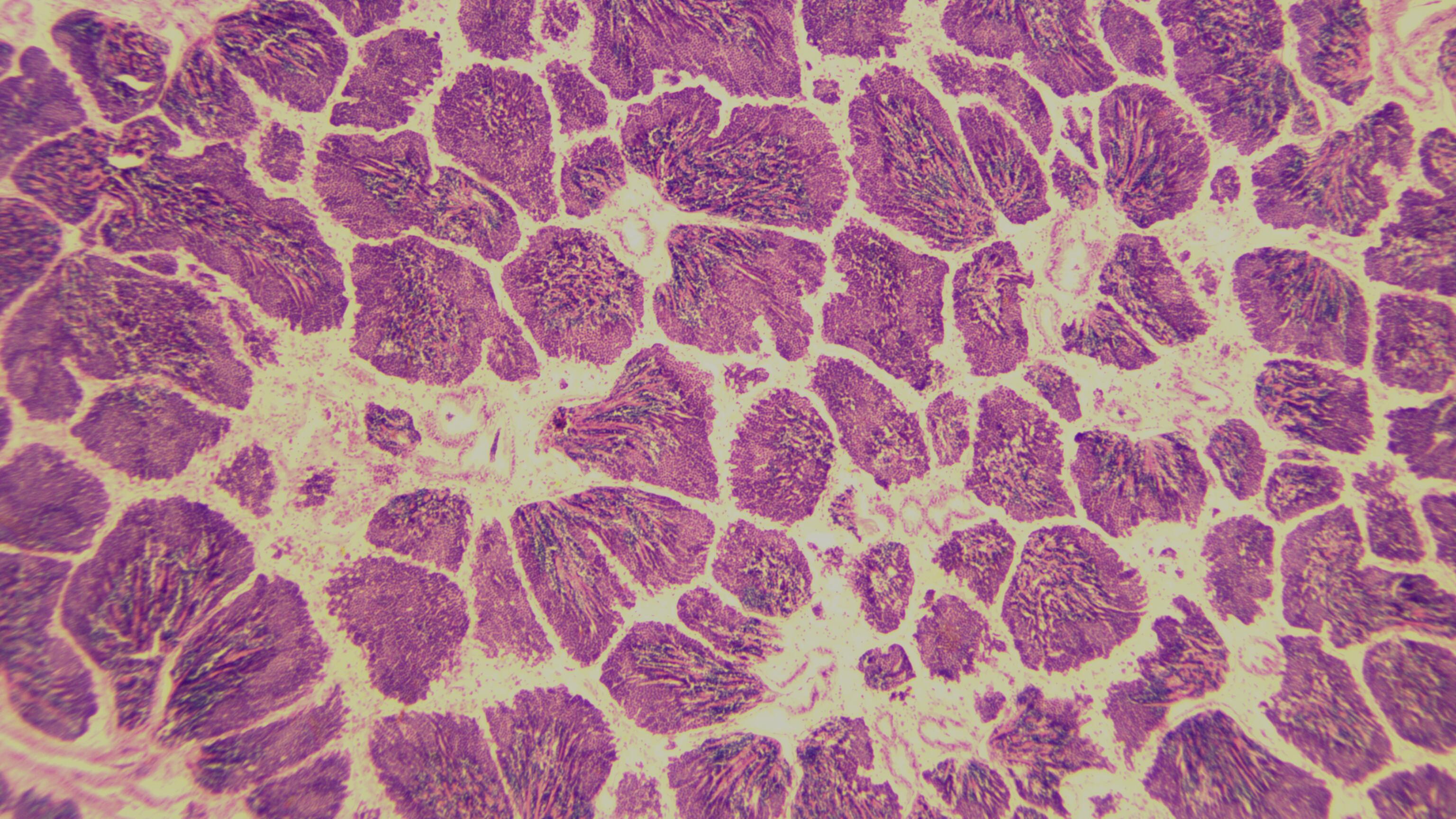
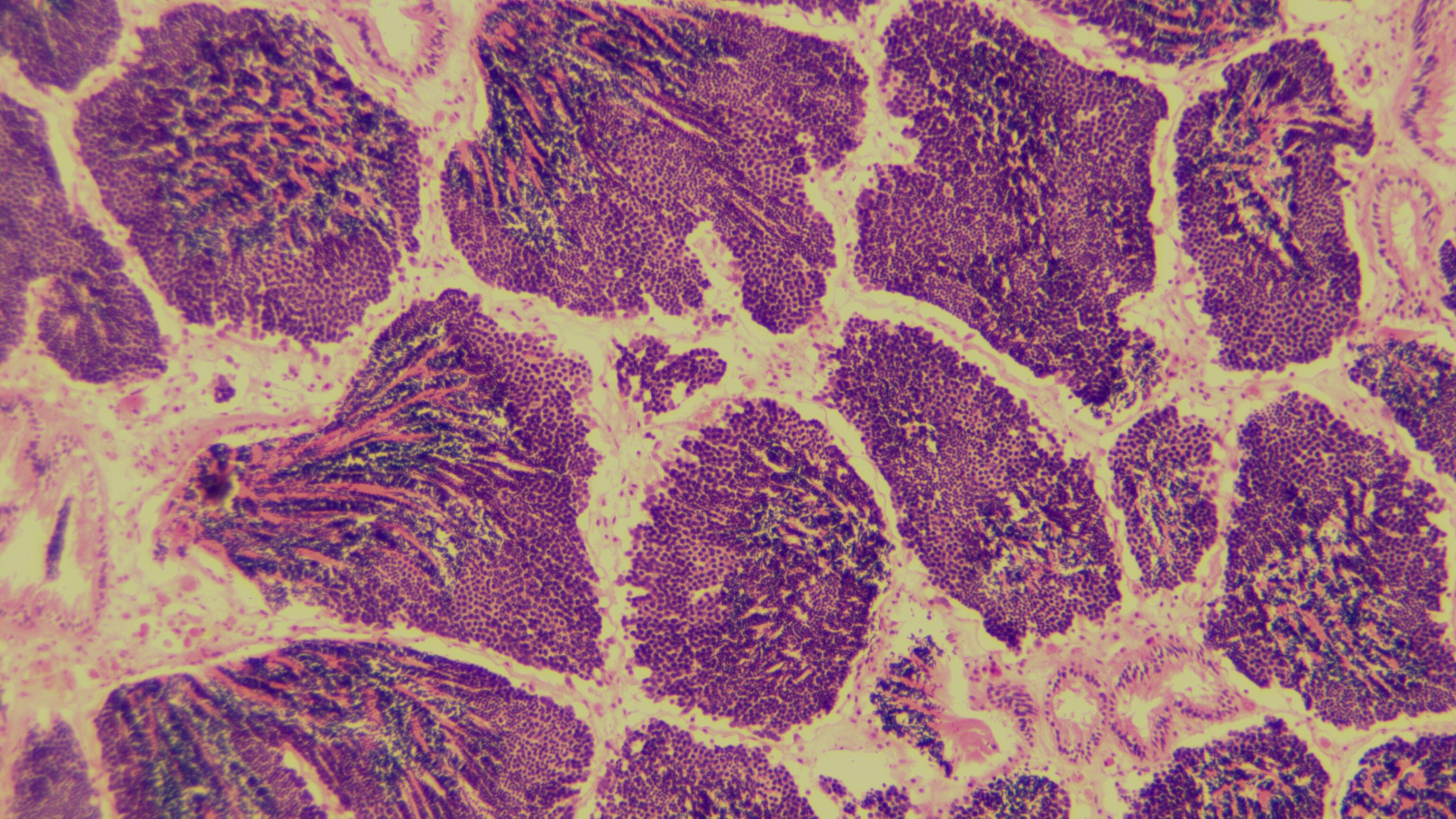
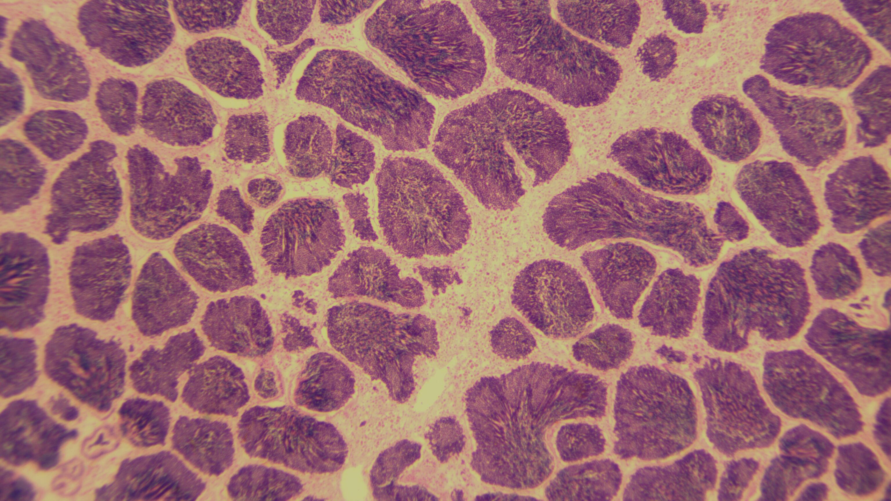
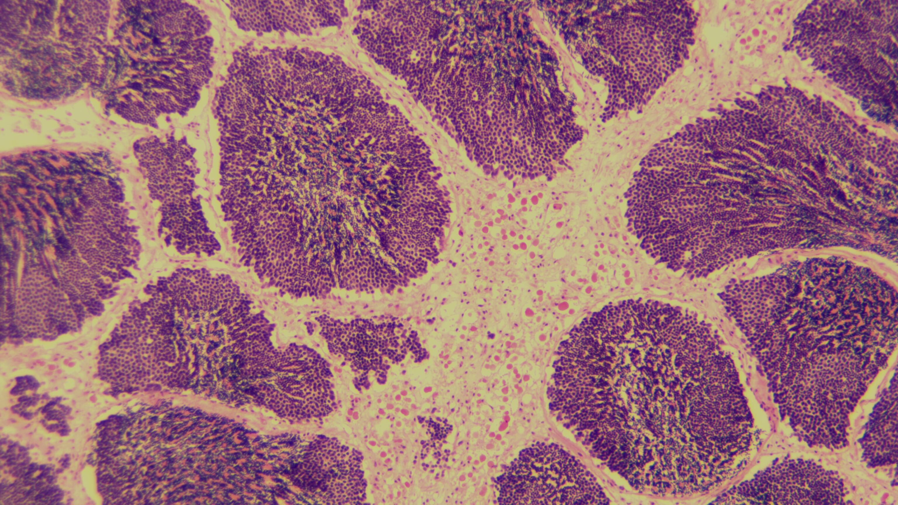
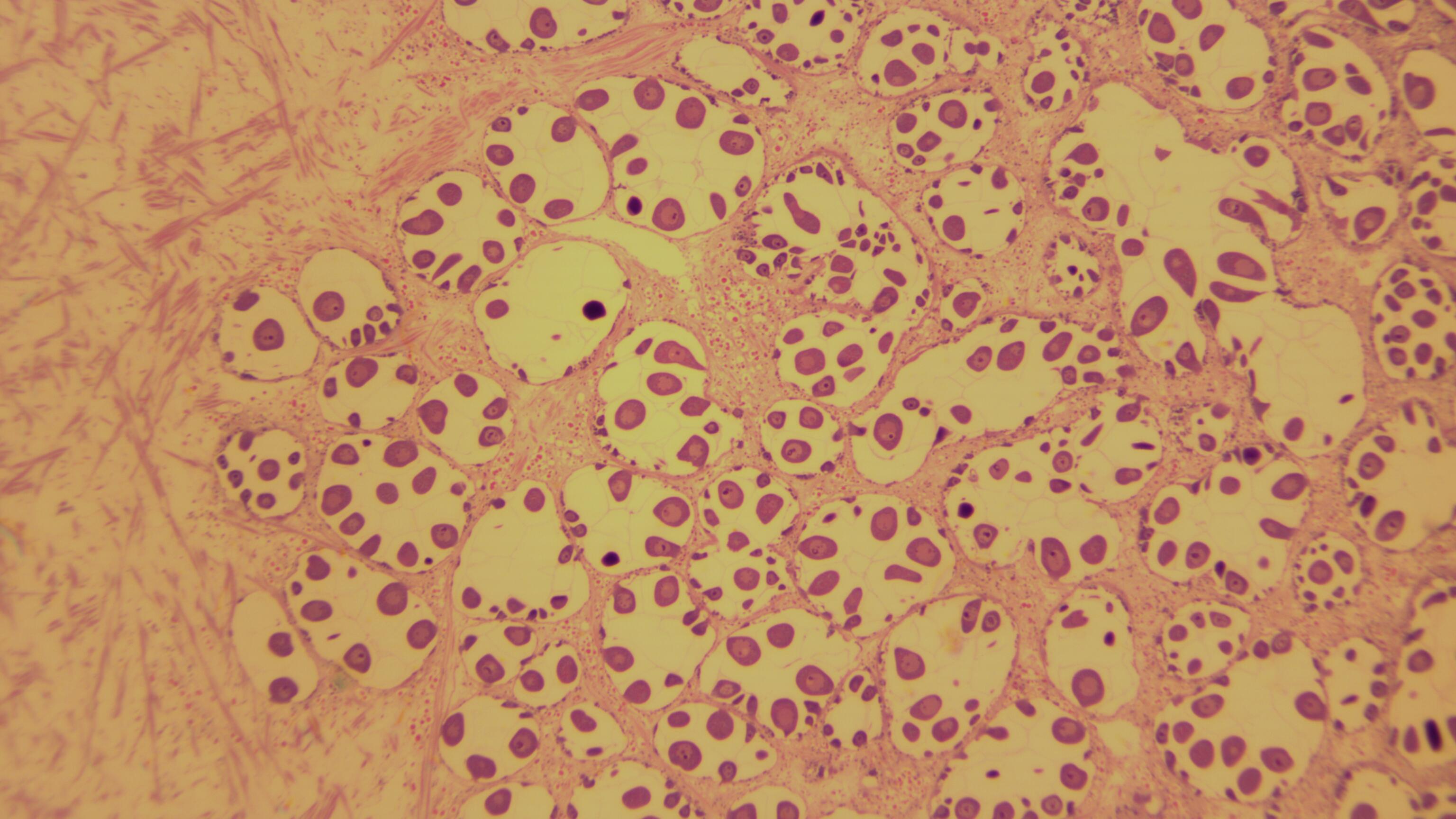
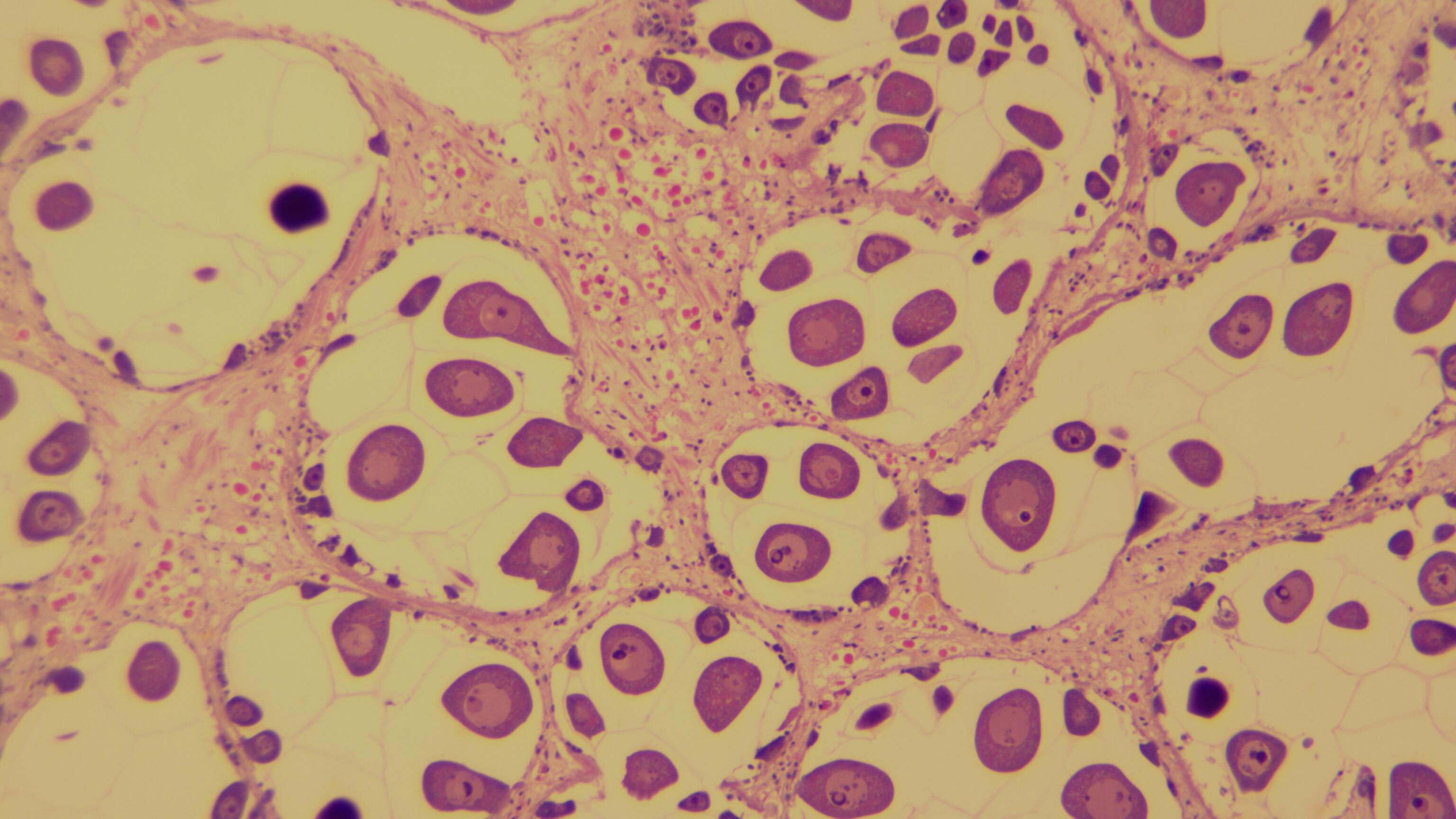
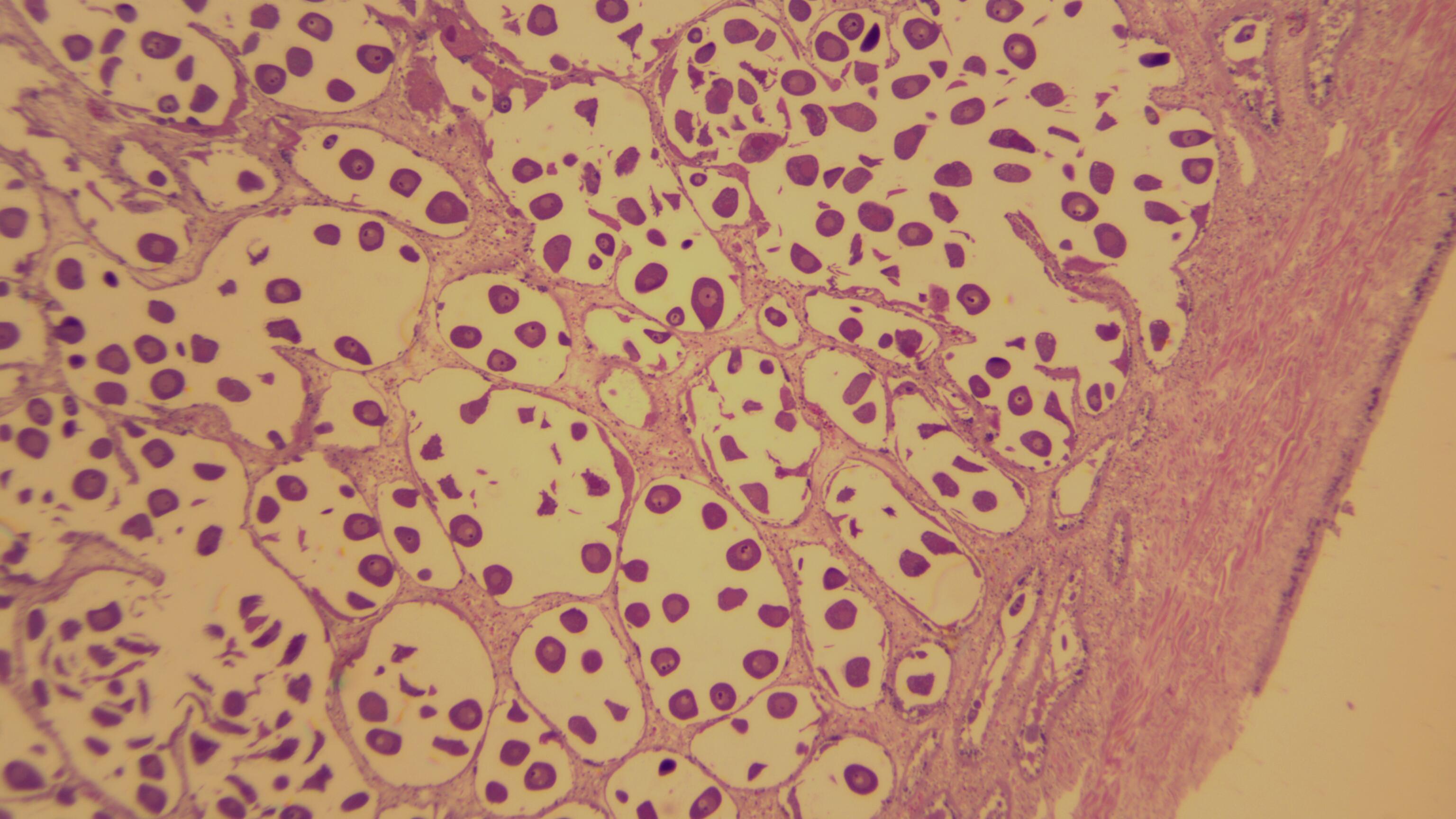
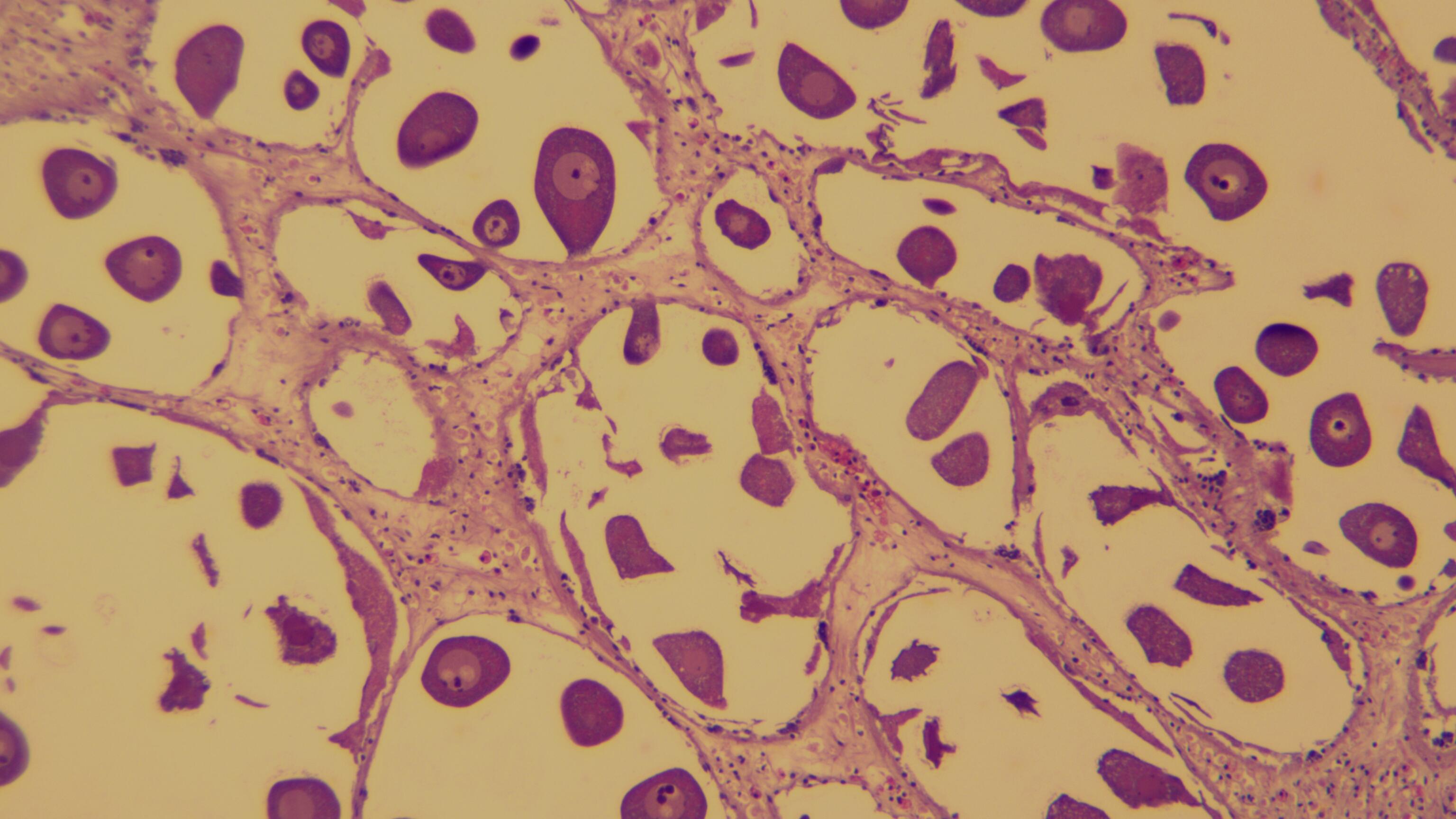
Received the histology blocks and H&E-stained slides for the Panopea generosa (Pacific geoduck) (Wikipedia) gonad samples submitted on 20260414 (Notebook entry). Slides were added to the histology database (Google Sheet).
Additionally, I imaged the slides at 4x and 10x objective magnications. Image files are named like so:
20260427-1-A-X-4x-01.tif where: - 20260427 is the date (April 27, 2026) - 1-A-X is the sample ID - 4x is the magnification - 01 is the image number
Slides were given to Kendall Barton (Marine Ecologist) of the Tulalip Tribes on 20260519 to be able to analyze them in more detail.
All images are available here:
MATERIALS & METHODS
Slides were imaged using using an Olypmus BH2-RFCA microscope with an AmScope MU530-BI camera. Images were captured using AmLite software and saved as TIFF files. Files also converted to JPEG format for easier viewing and sharing. Each slide was imaged at 4x and 10x magnification, with a single image taken at each magnification.
More specifically, the process for imaging the slides was as follows:
Place the slide on the microscope stage.
Start with the 4x objective lens and focus on the tissue section.
Capture the image using the AmLite software and save it as a TIFF file.
- Select
MU530-BIas the camera. - Click the
Snapbutton to capture the image. - Click on the captured image (tabbed next to live image tab) to open it in the image viewer.
- Click the
Savebutton in the upper left corner of the software to save the image as a TIFF file, using the naming convention described above.
- Select
Switch to the 10x objective lens and refocus on the same area of the tissue section.
Repeat steps 3a-3d to capture and save the image at 10x magnification.
Capture the image at 10x magnification and save it as a TIFF file.
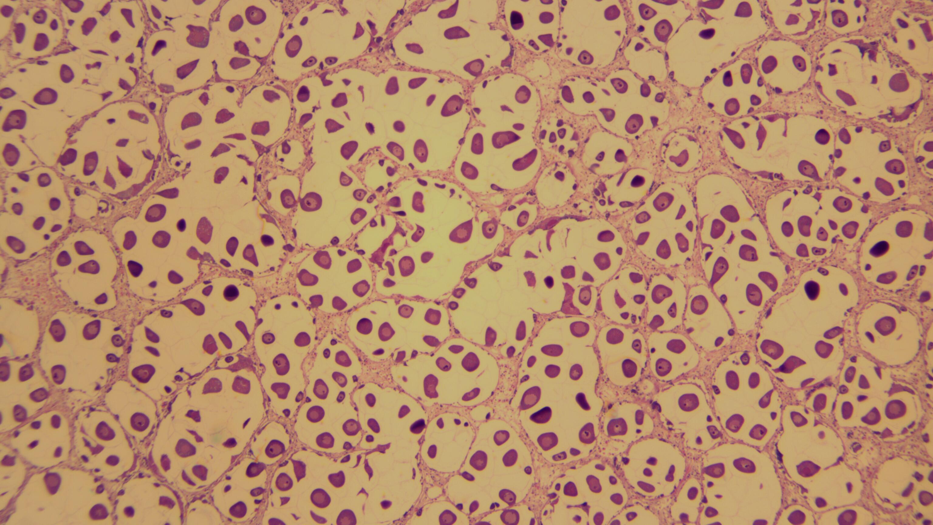
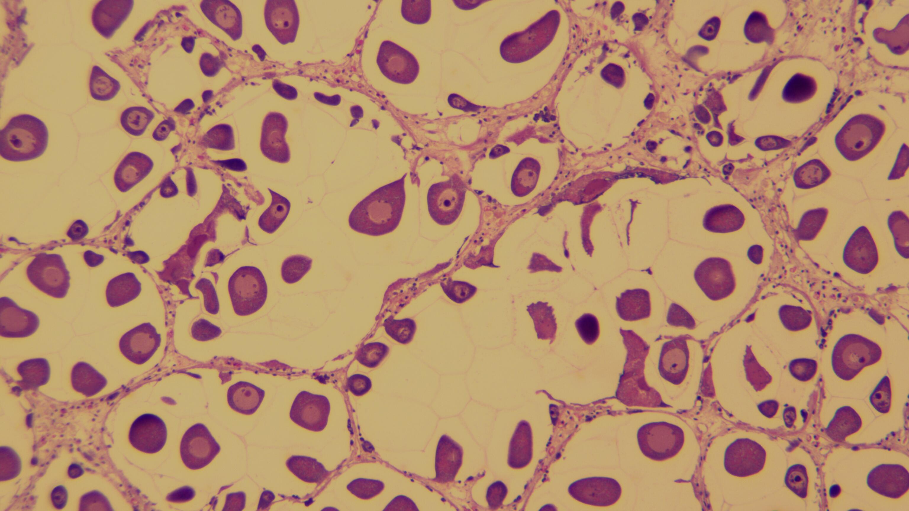
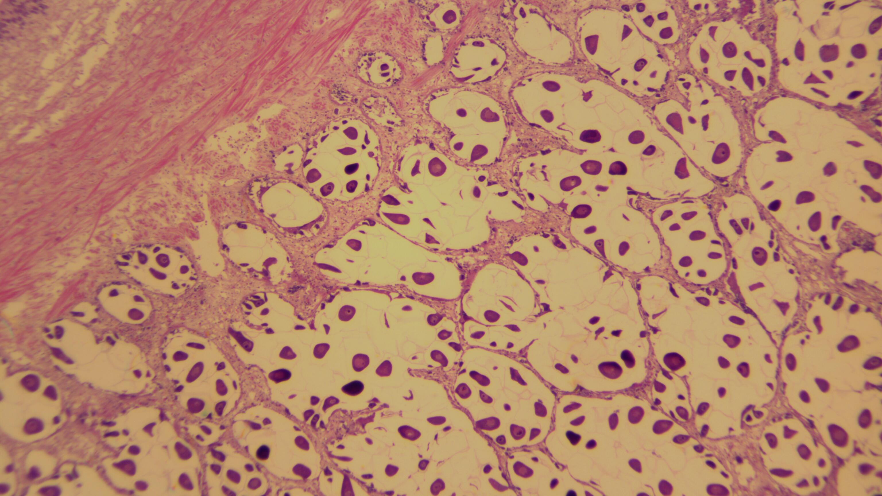
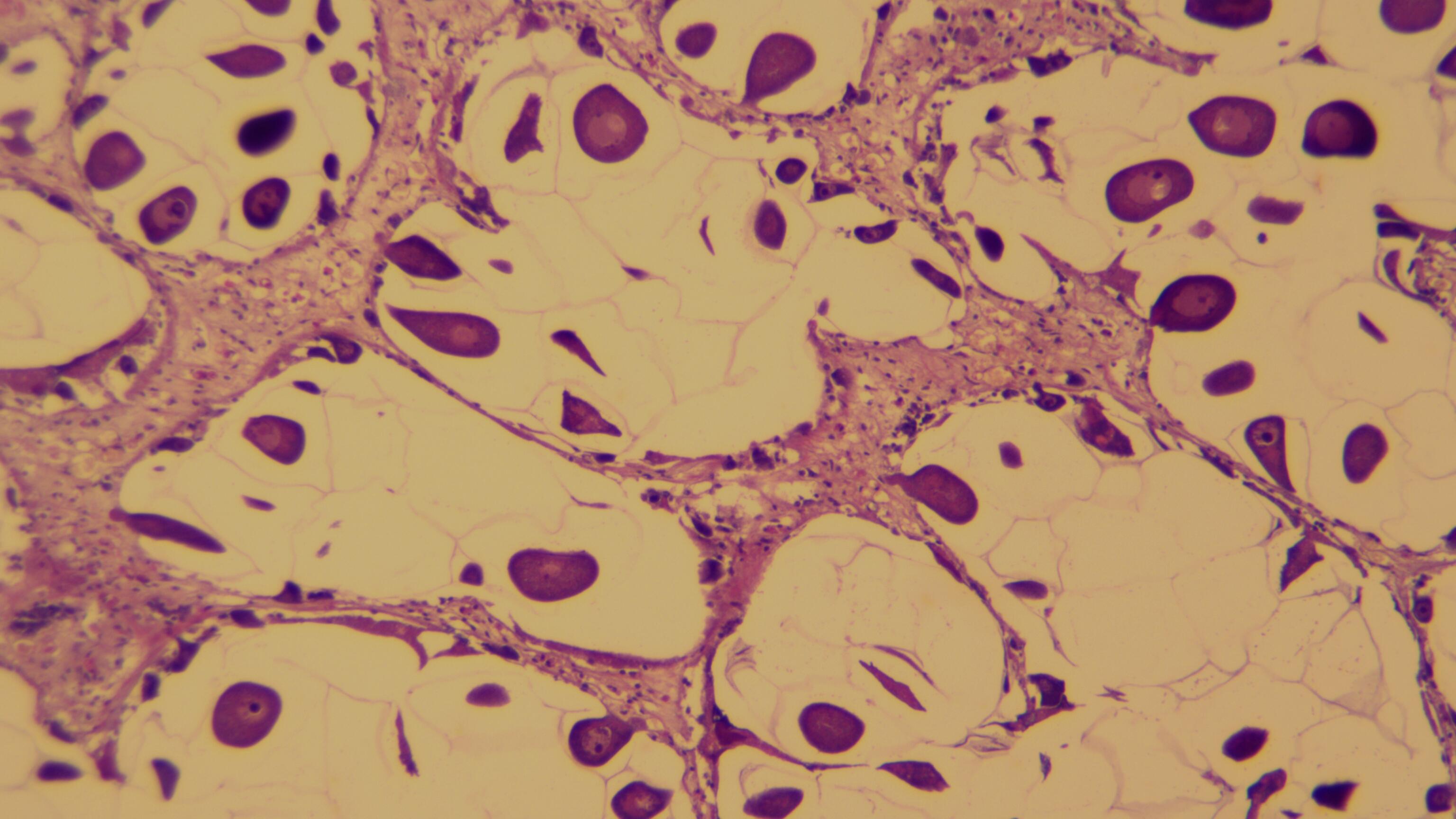
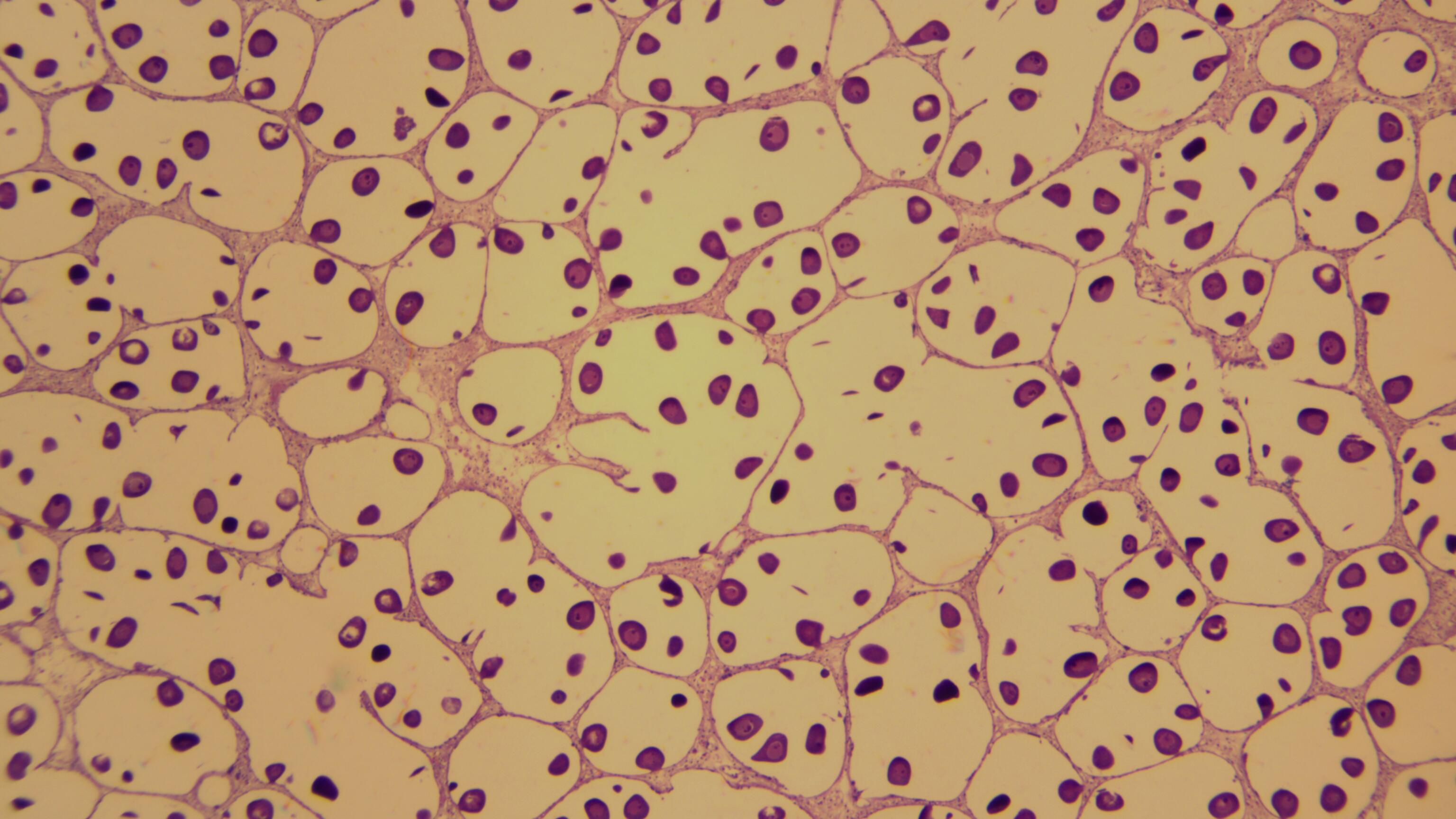
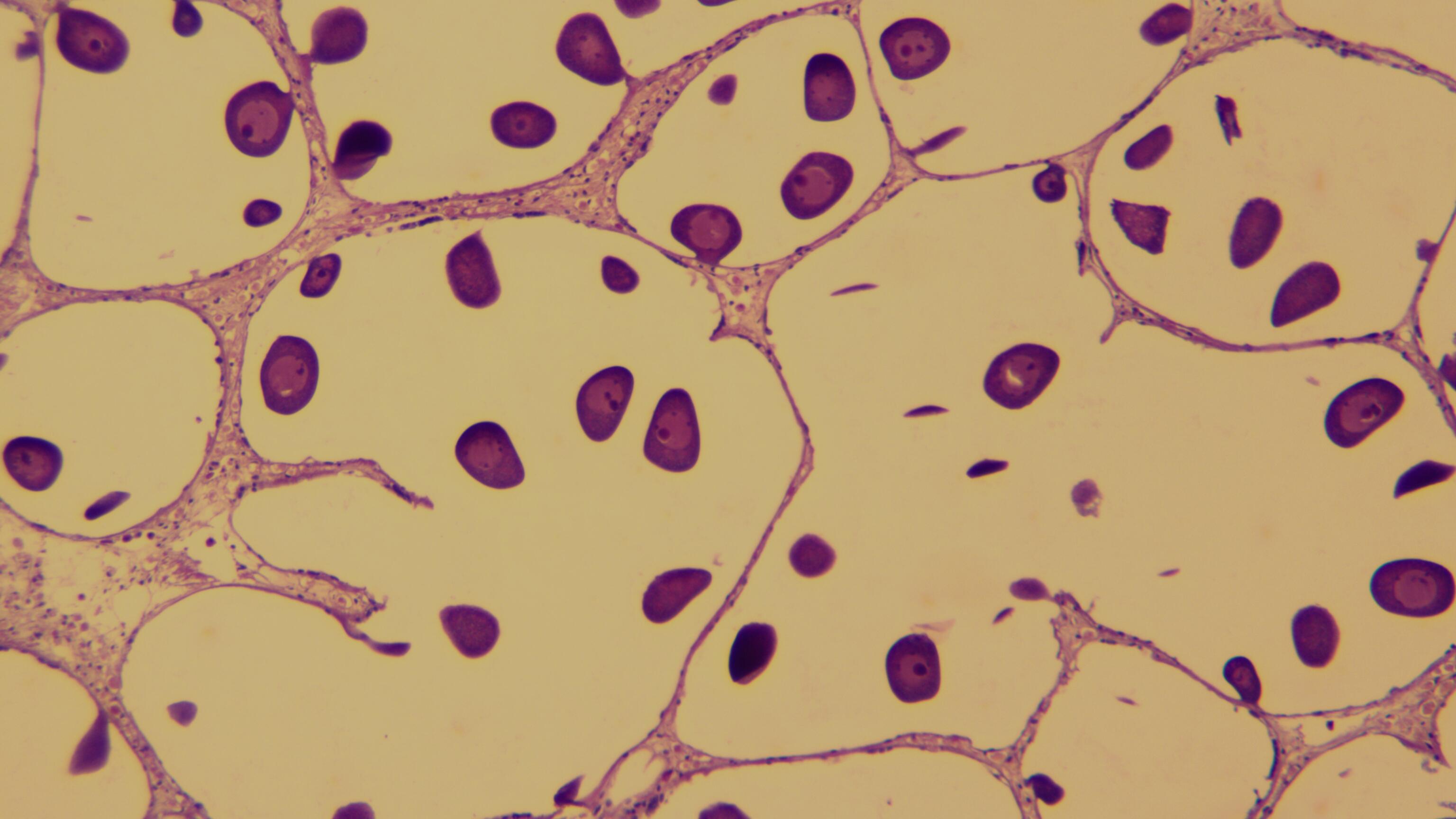
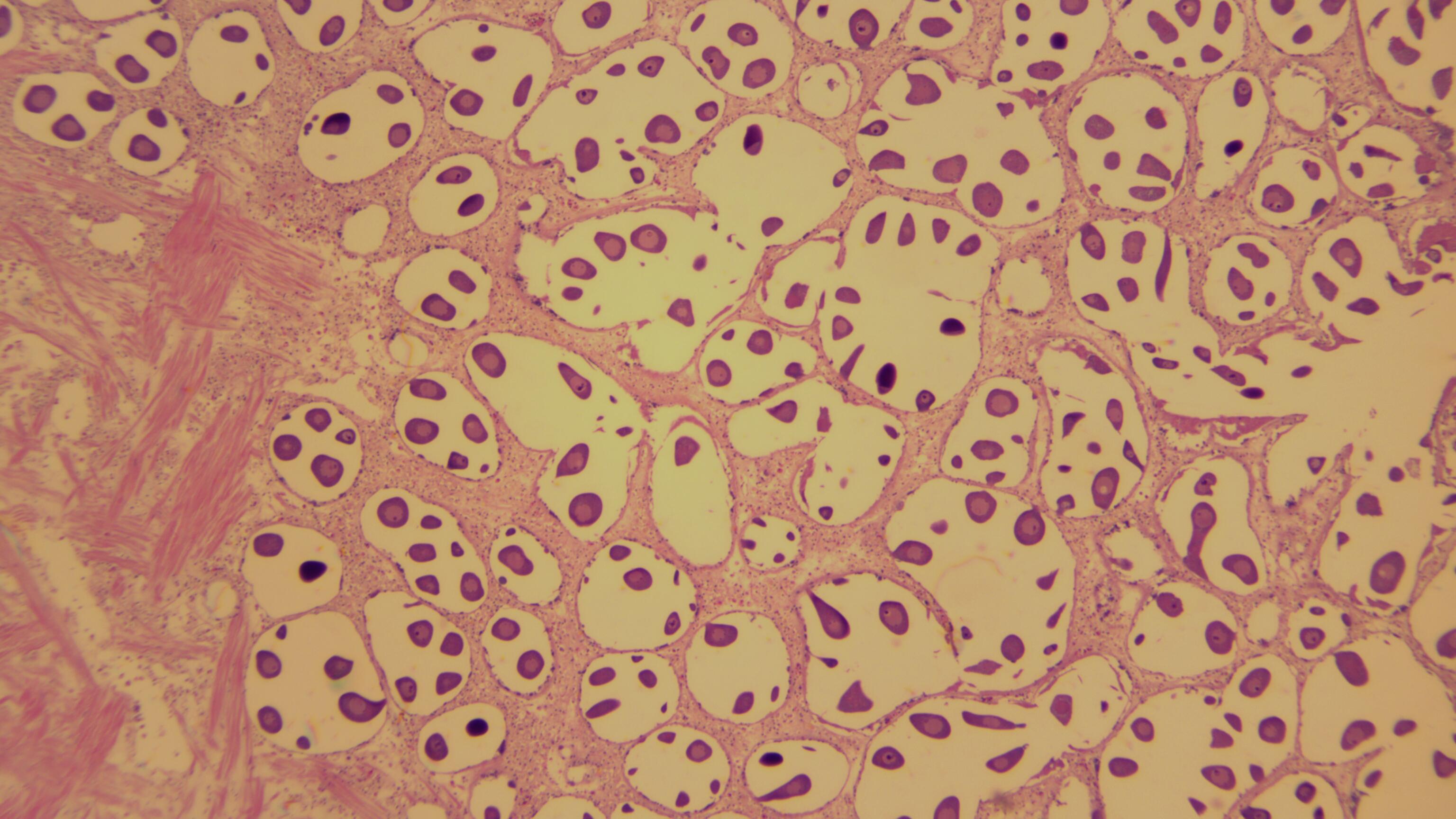
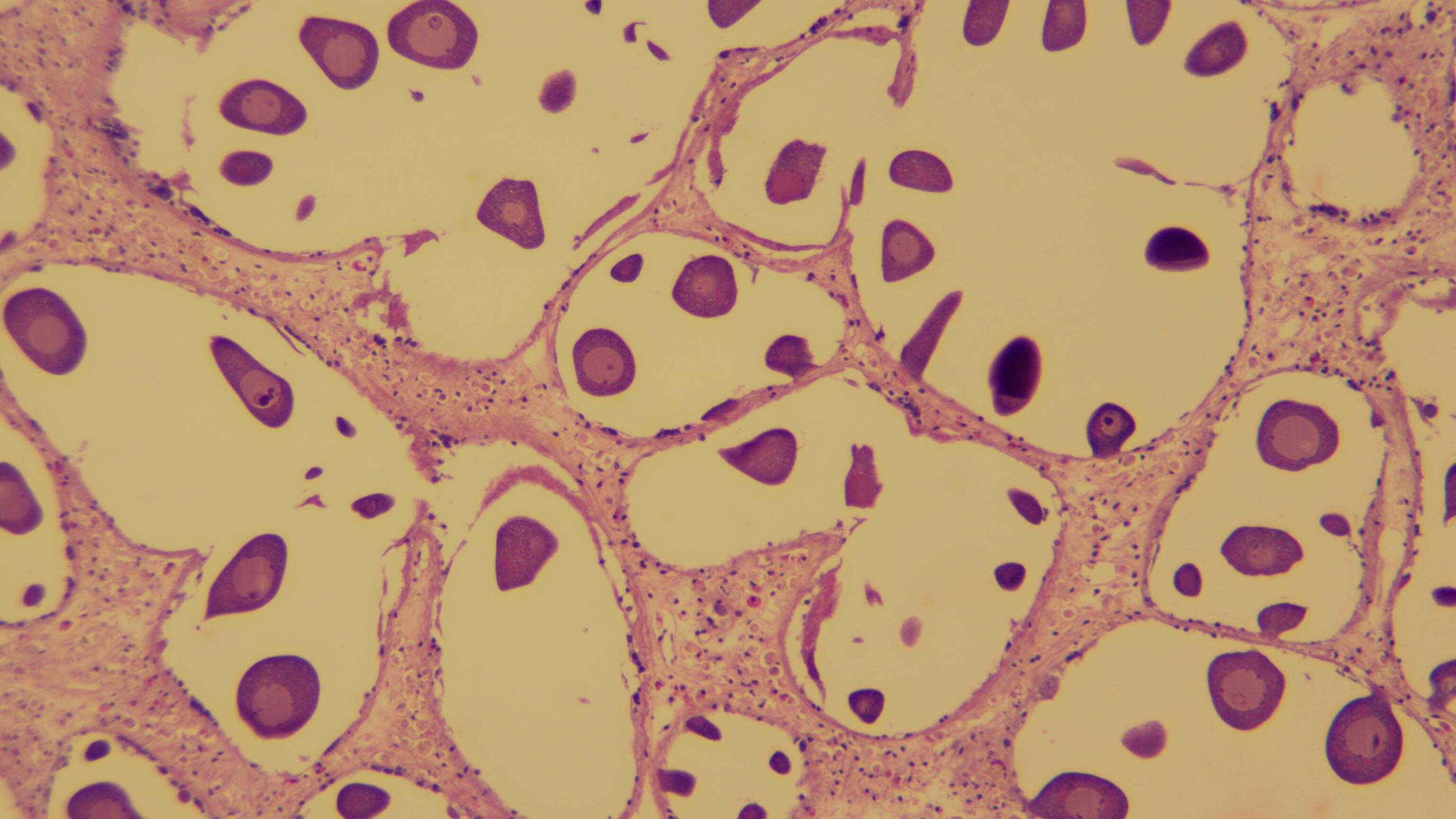
RESULTS
All images are below.